News & Events
VENTRICULAR TACHYCARDIA ABLATION USING 3D ELECTRO ANATOMIC MAPPING FOR THE FIRST TIME IN MALABAR
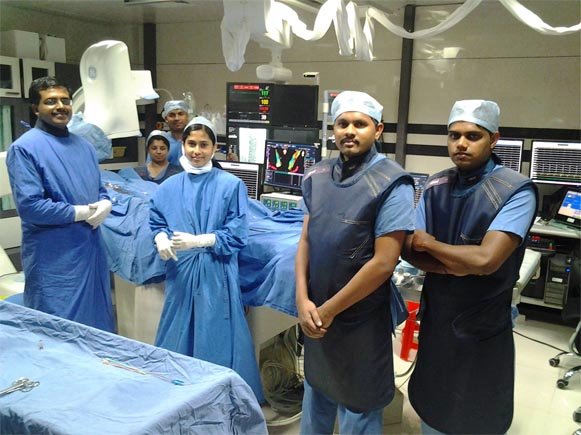
Sep 17, 2016
For the first time in Malabar, THREE cases of Ventricular Tachycardia were ablated using NAV X 3D electro anatomic mapping system at MICC. The procedure was done by Dr. ARUN GOPI, Consultant Cardiologist and Electrophysiologist. Ventricular tachycardia (VT) is an abnormal rapid heart rhythm originating from the lower pumping chambers of the heart (ventricles). Abnormal and fast rhythms from the ventricle may impair the ability of the pump to supply blood to the brain and the rest of the body. This may result in palpitations (a feeling of rapid or abnormal heart beat), dizziness, or syncope (loss of consciousness). If the heart rate increases to more than 300 beats per minute and becomes totally uncoordinated, called ventricular fibrillation (VF), sudden cardiac death will ensue. In fact VT/VF is the most common cause of sudden death in cardiac patients. There are 3 treatment options for VT: an ICD, antiarrhythmic medications, or catheter ablation, although many patients require a combination. In those who are still symptomatic on drugs the only curative treatment is radiofrequency ablation. VT ablation using the conventional mapping system has low success rate compared to 3D electroanatomic mapping. Electroanatomic mapping system is a modern electrophysiological mapping system that allows superposition of electrophysiologic data and cardiac anatomy. This results in three-dimensional reconstruction of mapped heart chamber together with color-coded activation sequence and/or voltage. The system allows precise orientation within mapped cavity and design of ablation lines between two anatomical barriers. In comparison with conventional mapping technique the success rate of catheter ablations increases while radiation exposure for patient and staff remains low. It is a minimally invasive procedure usually done under local anesthesia.